Uvea Clinic
What is Uveitis
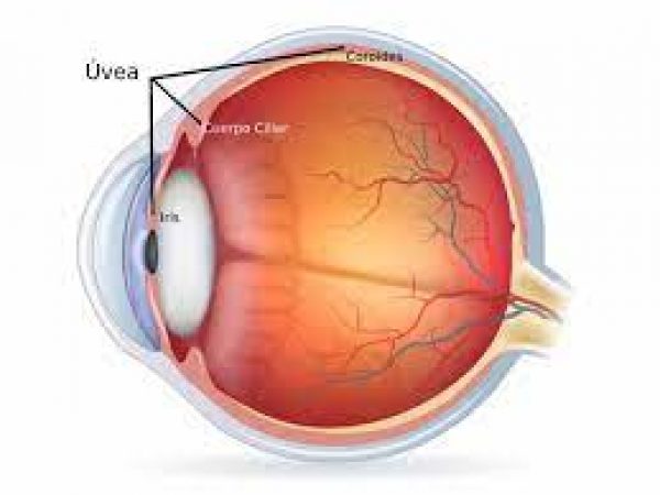
The uvea is the middle layer of the eye which contains much of the eye’s blood vessels. It is located between the sclera, the eye’s white outer coat, and the inner layer of the eye, called the retina, and is further made up of the iris, ciliary body, and choroid.
Uveitis encompasses a group of inflammatory diseases that produces swelling of the uveal tissues. It is not necessarily limited to the uvea but can also affect the lens, retina, optic nerve, and vitreous, producing reduced vision or blindness.
Uveitis may be caused by problems or diseases occurring in the eye or it can be part of an inflammatory disease affecting other parts of the body.
It can happen at all ages and primarily affects people between 20-60 years old.
Uveitis can last for a short (acute) or a long (chronic) time. The severest forms of uveitis can reoccur many times.
Symptoms
Uveitis can affect one or both eyes simultaneously. Symptoms may develop rapidly and can include:
- Blurred vision
- Dark, floating spots/lines in the vision (floaters)
- Eye pain
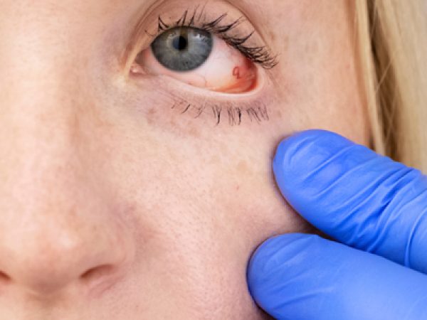
- Redness of the eye
- Sensitivity to light (photophobia)
The signs and symptoms of uveitis depend on the type of inflammation.
Acute anterior uveitis may occur in one or both eyes and in adults is characterized by eye pain, blurred vision, sensitivity to light and redness.
Intermediate uveitis causes blurred vision and floaters. Usually it is not associated with pain.
Posterior uveitis can produce vision loss. This type of uveitis can only be detected during an eye examination.
Causes
Inflammation is the body’s natural response to tissue damage, germs, or toxins. It produces swelling, redness, heat, and destroys tissues as certain white blood cells rush to the affected part of the body to contain or eliminate the insult.Any inflammation to the uveal tissue produces Uveitis.
Uveitis may be caused by:
- An attack from the body’s own immune system (autoimmunity)
- Infections or tumors occurring within the eye or in other parts of the body
- Trauma to the eye
- Drugs and toxins
- Most of the times the cause remains unknown which is termed as idiopathic
What are the types of Uveitis?
The type of uveitis can classified by where inflammation occurs in the uvea:
- Anterior uveitis is inflammation of the iris (iritis) or the iris and ciliary body.
- Intermediate uveitis is inflammation of the ciliary body.
- Posterior uveitis is inflammation of the choroid.
- Diffuse uveitis (also called pan-uveitis) is inflammation of all parts of the uvea.
How will my eye doctor check for Uveitis?
Diagnosis of uveitis includes a thorough patient’s medical history and a detailed examination of the eye to record the findings.
Further ancillary investigations , laboratory tests may be done to rule out an infection or an autoimmune disorder.
Eye examination includes
An Eye Chart or Visual Acuity Test: This test measures whether a patient’s vision has decreased.
AOcular Pressure
AA Slit Lamp Exam: A slit lamp noninvasively inspects the front and back parts of the eye
AA Dilated Fundus Examination: The pupil is widened (dilated) with eye drops and then a light is shown through with an instrument called an ophthalmoscope to noninvasively inspect the back, inside part of the eye.
AWhat are the complications of Uveitis?
Many cases of uveitis are chronic, and they can produce numerous possible complications, including clouding of the cornea, cataracts, elevated eye pressure (IOP), glaucoma, swelling of the retina or retinal detachment. These complications can result in permanent vision loss.
AWhat’s the treatment for Uveitis?
The goal of treatment in uveitis is to eliminate inflammation, alleviate pain, prevent further tissue damage, and restore any loss of vision.
If uveitis is caused by an underlying condition, treatment will focus on that specific condition.
Drugs that reduce inflammation. Your doctor may first prescribe eyedrops with an anti-inflammatory medication, such as a corticosteroid. If those don’t help, a corticosteroid tablets or injection may be the next step.
Drugs that fight bacteria or viruses. If uveitis is caused by an infection, your doctor may prescribe antibiotics, antiviral medications or other medicines, with or without corticosteroids, to bring the infection under control.
Drugs that affect the immune system or destroy cells. You may need immunosuppressive or cytotoxic drugs if your uveitis affects both eyes, doesn’t respond well to corticosteroids or becomes severe enough to threaten your vision.
ASurgical and other procedures
Vitrectomy. Surgery to remove some of the vitreous in your eye (vitrectomy) may be necessary to manage the condition.
Surgery that implants a device into the eye to provide a slow and sustained release of a medication. For people with difficult-to-treat posterior uveitis, a device that’s implanted in the eye may be an option. This device slowly releases corticosteroid medication into the eye for two to three years. Possible side effects of this treatment include cataracts and glaucoma.
AAnterior Uveitis treatments
Anterior uveitis may be treated by:- Taking eye drops that dilate the pupil to prevent muscle spasms in the iris and ciliary body (see diagram)
- Taking eye drops containing steroids, such as prednisone, to reduce inflammation
AIntermediate, Posterior, Panuveitis treatments
Intermediate, posterior, and panuveitis are often treated with injections around the eye, medications given by mouth, or, in some instances, time-release capsules that are surgically implanted inside the eye. Other immunosuppressive agents may be given. A doctor must make sure a patient is not fighting an infection before proceeding with these therapies.
Some of these medications can have serious side effects, such as glaucoma and cataracts. You may need to visit your doctor for follow-up examinations and blood tests every 1 to 3 months.


